

Case Report
Successfully treated Loa Loa meningitis in a patient domiciled in Central Africa Republic
1Neurology department Mohammed V Military Instruction University hospital. Mohammed V University Souissi. Faculty of medicine and pharmacy of Rabat. Morocco.
2Anatomy department. Mohammed V Souissi universities. Faculty of medicine and pharmacy of Rabat. Morocco.
*Corresponding Author: Jawad Oumerzouk., Neurology department Mohammed V Military Instruction University hospital. Mohammed V University Souissi. Faculty of medicine and pharmacy of Rabat. Morocco.
Citation: Oumerzouk J., Hafid J., Bourazza A. (2026). Successfully treated Loa Loa meningitis in a patient domiciled in Central Africa Republic, Clinical Case Reports and Studies, BioRes Scientia Publishers. 13(1):1-4. DOI: 10.59657/2837-2565.brs.26.338
Copyright: © 2026 Jawad Oumerzouk, this is an open-access article distributed under the terms of the Creative Commons Attribution License, which permits unrestricted use, distribution, and reproduction in any medium, provided the original author and source are credited.
Received: July 14, 2026 | Accepted: July 28, 2026 | Published: August 04, 2026
Abstract
Neurological complications of direct central nervous system (CNS) involvement during Loa Loa infection present real danger. We herein present a case of a 27-year-old man domiciled in Central African Republic Lo presenting a Loa meningitis unrelated to ant filarial treatment who was successfully treated with ivermectin. The parasitology examination of a peripheral blood sample revealed the presence of Loa loa microfilariae with a low parasitemia of 100 parasites per microliter and numerous live microfilariae were seen on cerebrospinal fluid (CSF) microscopy with estimated count of 20 microfilariae /50 microliter. Giemsa and Haematoxylin stain demonstrated sheathed microfilariae with nuclei extending to the tip of the larva worm. Our patient received a single oral dose of 9 mg of ivermectin (1.5 tablets of 6 mg), and the patient's neurological condition improved. A follow-up CSF analysis 2 weeks later showed the absence of Loa loa microfilariae in both body fluids.
In conclusion, even in the absence of previous antifilarial treatment, Loa Loa microfilariae can cause the blood-brain barrier disruption and penetrate into the CNS, CSF and meningeal space where they may induce meningitis or meningoencephalitis. Therefore, filarial infections should be suspected in the setting of neurological manifestations in endemic regions, especially in the presence of other infections such as malaria.
Keywords: Loa Loa, meningitis, cerebrospinal, ivermectin, parasitology
Introduction
Loiasis is a vector -borne parasitic infection transmitted by the filarial nematode Loa loa of the genus Chrysops [1]. This disease es endemic across many areas of Central and West Africa, particularly rainforest areas [1]. However, imported cases are described throughout the world, due to intense economic and touristic population exchanges [2]. It is estimated that 30 million people are at risk of acquiring loiasis, while over 10 million individuals living in endemic zones are thought to be already infected. However, this prevalence based on the detection of microfilariae in blood, it seems to be underestimated, as about one- third of subjects are amicrofilaremic [3].
Additionally, adult worms living in subcutaneous tissues and in the fascial layers produces microfilariae circulating in peripheral blood and both parasite stages may invade any location in the human body, to produce a huge variety of clinical manifestations [1]. However, n eurological manifestations of direct central nervous system (CNS) involvement present real danger [1]. We herein present a case of successfully treated Loa Loa meningitis unrelated to antifilarial treatment.
Case Report
A 27-year-old man domiciled in Central African Republic, a region endemic for loiasis who had been treated in 2009 for Plasmodium falciparum malaria, presented with 8 days history of persistent, band-like headaches, without seizures, intracranial hypertension or consciousness disorders. The patient was totally asymptomatic prior to illness and there was no medical history of hypertension, diabetes or drug ingestion. Patient denied history of itching, swelling of the body, eye pain or history of antifilarial drugs (ivermectin or diethylcarbamazine) within the days preceding his hospitalization.
The essential findings on clinical examination were fever (temperature 38°C) and neck stiffness. The patient's vital signs were within normal limits; blood pressure was 120/60 mmHg, the pulse rate was 78 beats per minute, SpO2=98%, his Glasgow Coma Scale (GCS) was 15/15, pupils were equal and reactive bilaterally, and his Motor power was 5/5 in his upper and lower limbs. A fundus examination was essentially normal and we could not see or recover adult worm on probing the subconjunctival tissue. In our patient, skin involvement, ocular lesions, Calabar edema and pruritus were not found.
The blood tests showed anemia and eosinophilia (table), leading us to suspect a parasitic infection. The thick and thin blood film was negative for malaria parasite and the liver function tests performed the same day were normal. The parasitology examination of a peripheral blood sample revealed the presence of Loa loa microfilariae with a parasitemia of 100 parasites per microliter (figure 1). Screening tests for humans’ immunodeficiency virus (HIV) infection, hepatitis B and C viruses, and syphilis respectively were Negative and radiological investigations (MRI brain) were normal. To lumbar puncture performed, revealed a clear and colorful cerebrospinal fluid (CSF) with normal CSF opening pressure and slightly increased white blood cell count (table 1). The CSF protein and sugar they were normal. Direct bacteriological examination of the CSF was negative, and numerous live microfilariae were seen on CSF microscopy with estimated count of 20 microfilariae /50 microliter. Giemsa and Haematoxylin stain demonstrated sheathed microfilariae with nuclei extending to the tip of the larva worm. The CSF and blood cultures for bacterial or fungal infections were persistently negative and china ink stain to detect Cryptococcus neoformans capsules was also negative.
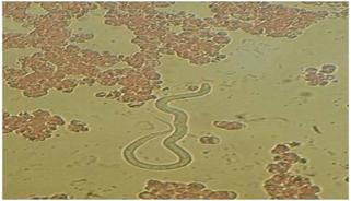
Figure 1: The parasitology examination of a peripheral blood sample revealed the presence of Loa loa with a parasitemia of 100 parasites per microliter.
Table 1: Blood and cerebrospinal fluid characteristics in our patient presenting Loa Loa meningitis
| Biological tests | Value | Reference range | |
| Hemoglobin (g/ dL) | 7.6 g/ dL | 11-16 | |
| White blood cell count (cells /mm³) | 9300 | 4000-10000 | |
| Eosinophils (cells /mm³) | 1,400 (15.4%) | 46-630 | |
| CSF opening pressure (mmHg) | 10 | <18> | |
| CSF cytology | White blood cell count (WBC/mm3) | 11 | 0-5 |
| Red blood cell count (RBC/mm3) | 3 | 0-5 | |
| Biochemical characteristics | CSF protein (g/L) | 0.30 | 0.15-0.45 |
| CSF sugar (g/L) | 0.65 | 0.40-0.70 | |
CSF: Cerebrospinal fluid
With our clinical examination and investigations excluding other possible causes of meningitis, the Loa loa microfilariae presence in the CSF of this patient was the primary mechanism responsible for the meningitis rather than a secondary event. Due to his low parasitemia (low Loa loa) filaria count of 100 parasites per microliter), our patient received a single oral dose of 9 mg of ivermectin (1.5 tablets of 6 mg), and there were no neurological complications related to this therapy. The patient's neurological condition improved with resolution of his headaches and eosinophilia. A lumbar follow-up puncture revealed a white blood cell count of 1/mm³ and the repeated blood film and CSF fluid analysis 2 weeks later showed the absence of Loa loa microfilariae in both body fluids.
Discussion
The classical signs of the chronic Loa Loa infection are itching, unpleasant sensation of migratory movements of the adult worms under the conjunctiva and Calabar swellings [1,2]. The neurological complications associated with Loa loa infection are usually the consequence of treatment with diethylcarbamizine (DEC) or ivermectin (Mectizan), although Item may be rarely caused spontaneously by adult worm or of microfilaria (mf) [3,4,5,6]. Indeed, even in the absence of previous antifilarial treatment, Loa Loa microfilariae can cause the blood-brain barrier (BBB) disruption and penetrate into the CNS, CSF and meningeal space where they may induce cellular immunological reaction [7,8,9]. This immune response could play a crucial role through proinflammatory cytokines and vasoactive amines release, complement activation and influx of inflammatory cells causing vessel destruction, meningitis or meningoencephalitis [3,4,7]. Additionally, the chronic circulation of mf may also lead to cerebral microcirculation obstruction or hemorrhages[8]. Therefore, the neurological manifestations may include asthenia, somnolence, headaches, motor and/or sensory deficits, troubles of sensation, brainstem syndrome, cognitive impairment and altered consciousness [2,7]. Nevertheless, that neurological complications depend upon many factors such as the levels of circulating Loa loa microfilariae in the bloodstream (typically exceeding ≥ 30,000 microfilariae / mL), duration and level of exposure, number of secondary bacterial and fungal infections, the degree of host immune response and others cofactors such as drugs and coinfection [ 1]. Tour knowledge, the only cases of meningoencephalitis unrelated to antifilarial drug in infected patients with Loa loa are those reported by Vitris M et al (1989)., and Taiwo SS et al (2007) and in these 2 cases, the disease course was unfavorable with fatal outcome [5,6]. Thus, we have described in this article an original case of meningtis associated with Loa Loa infection in a patient harboring low parasitemia, who was successfully treated with a single oral dose of 9 mg of ivermectin.
The first cases of threatening neurological serious adverse event (SAEs) following the use of antifilarial treatment for loiasis began to be described in regions that were co-endemic for Loa Loa and Onchocerca volvulus (river blindness) with significantly higher risk when the Loa loa microfilaremia exceed 8000 microfilariae per mL of blood [4,8,9]. Thus, it It is probable that the drug provokes the passage of Loa Loa microfilariae into the CSF and brain tissue with a massive mf death, and that the latter, floating passively in the blood circulation, may finally provoke embolisms in the capillaries and induces inflammation, especially in the brain (encephalopathies) [5,6]. This passage may be facilitated by some conditions including concomitant infections (trypanosomiasis, syphilis, onchocerciasis, acute encephalitis, or Plasmodium infection); comorbidities chronic vascular disease) and genetic predisposition [1,2,5,6,8]. Therefore, in order to reduce the incidence of DEC- related neurologic SAEs in subjects with high pre-treatment level of microfilaremia (≥8000 mf / mL), authors have recommended a 3-week regimen of albendazole (ALB), apheresis sessions, or ivermectin treatment [9].
According to 2003 Scientific Working Group on SAEs, the principles to follow for the management of Loa Loa ivermectin -related encephalopathy would appear to be early hospitalization, good nursing care, maintenance of fluid, electrolyte / nutritional support, antihistamines for putative inflammatory lesions in the CNS, and antibiotics to prevent or control possible secondary infections [3,4,8]. Corticosteroids should be avoided because evidence of efficacy es lacking, and potential of harmful effects [9].
Although regarded as a benign condition, significant excess Mortality has recently ascribed to the chronic infection by Loa Loa. Higher mortality and fatal outcome are essentially reported when CNS is directly involved and in cases with late diagnosis and delayed management [1,3]. Unfortunately, the majority of patients who have survived after spontaneous or antifilarial-induced Loa Loa encephalopathy showed sequellae including engine defects, extrapyramidal disorders, ataxia, disturbances of balance, aphasia, psychomotor slowing, psychiatric sequelae or dementia [6,9].
Conclusion
The spontaneous Neurological complications of Loa Loa infection are fairly rare and various. The risk of meningitis and encephalopathy in a previously healthy person occur essentially when the Loa loa microfilaremia exceed 8000 microfilariae per mL with the presence of Loa Loa mf in the CSF. We have described an original case of a patient presenting Loa Loa infection with low parasitemia and who developed meningitis unrelated to antifilarial drug, successfully treated with a single oral dose of 9 mg of ivermectin. Thus, such n eurological manifestations in endemic countries should raise the question of filarial infection and prompt early diagnosis and adequate treatment, to avoid severe sequellae.
Declarations
Funding declaration
No funding sources to declare
Conflict of interest
The authors have no conflict of interest
References
- Bhalla, D., Dumas, M., & Preux, P. M. (2013). Neurological manifestations of filarial infections. Handbook of Clinical Neurology, 114:235–242.
Publisher | Google Scholor - Taiwo, S. S., & Tamiowo, M. O. (2007). Loa loa meningoencephalitis in Southwestern Nigeria. West African Journal of Medicine, 26(2):156–159.
Publisher | Google Scholor - Moliner, J. V., Valverde, A. G., & Sorolla, J. M. (2011). Loa loa meningoencephalitis secondary to treatment with mebendazol. Medicina Clínica, 136(5):228–229.
Publisher | Google Scholor - Holmes, D. (2013). Loa loa: Neglected neurology and nematodes. The Lancet Neurology, 12(7):631–632.
Publisher | Google Scholor - Lukiana, T., Mandina, M., Situakibanza, N. H., et al. (2006). A possible case of spontaneous Loa loa encephalopathy associated with a glomerulopathy. Filaria Journal, 5:Article 6.
Publisher | Google Scholor - Checkouri, T., Missamou, F., Pion, S. D. S., et al. (2023). Association between altered cognition and Loa loa microfilaremia: First evidence from a cross-sectional study in a rural area of the Republic of Congo. PLoS Neglected Tropical Diseases, 17(6):e0011430.
Publisher | Google Scholor - Gobbi, F., Boussinesq, M., Mascarello, M., et al. (2011). Loiasis with peripheral nerve involvement and spleen lesions. The American Journal of Tropical Medicine and Hygiene, 84(5):733–737.
Publisher | Google Scholor - Boussinesq, M., Gardon, J., Gardon-Wendel, N., et al. (1998). Three probable cases of Loa loa encephalopathy following ivermectin treatment for onchocerciasis. The American Journal of Tropical Medicine and Hygiene, 58(4):461–469.
Publisher | Google Scholor - Volpicelli, L., De Angelis, M., Morano, A., et al. (2020). Encephalopathy in a patient with loiasis treated with albendazole: A case report. Parasitology International. Advance online publication.
Publisher | Google Scholor