

Research Article
New And Hitherto Unrecorded Black Mildew Causing Fungus on The Syzygium Mundagam an Endemic Plant in Southern Western Ghats, India
- Dr. Lini K. Mathew *
Assistant Professor & Research Guide, Postgraduate & Research Department of Botany, St. Thomas College, Kozhencherry, Kerala, India.
*Corresponding Author: Lini K. Mathew, Assistant Professor & Research Guide, Postgraduate & Research Department of Botany, St. Thomas College, Kozhencherry, Kerala, India.
Citation: Lini K. Mathew. (2025). New And Hitherto Unrecorded Black Mildew Causing Fungus on The Syzygium Mundagam an Endemic Plant in Southern Western Ghats, India, Journal of BioMed Research and Reports, BioRes Scientia Publishers. 8(3):1-4. DOI: 10.59657/2837-4681.brs.25.183
Copyright: © 2025 Lini K. Mathew, this is an open-access article distributed under the terms of the Creative Commons Attribution License, which permits unrestricted use, distribution, and reproduction in any medium, provided the original author and source are credited.
Received: June 10, 2025 | Accepted: June 24, 2025 | Published: July 01, 2025
Abstract
The foliicolous black mildew fungus infected the leaves of an endemic plant Syzygium mundagam. The present fungus was similar to the genus Asterina but differed from it in having septation at the extreme end of the ascospores making a significant difference in the size and shape of both cells. Hence, this fungus has been accommodated in the genus, Vishnumyces, which is described and illustrated in detail.
Keywords: foliicolous fungi; black mildew; new species
Introduction
During a survey of the foliicolous fungi in the Western Ghats region of Kerala state, author came across an endemic plant, Syzygium mundagam endemic to southern Western Ghats- South Sahyadri and between Palakkad hills to Coorg Region in Central Sahyadris (Henry et al., 1987, Gamble, 1997; Sasidharan, 2004), infected with a black mildew fungus. The microscopic analysis indicated that the fungus has a superficial mycelium, laterally situated appressoria, and thyriothecia that open stellately at the center, with the upper layer of the thyriothecia containing cells arranged radially. These characters confirms that the present fungus belongs to the family Asterinaceae (Arx and Muller, 1975). Notably, the septation at the terminal end of the ascospores distinguishes the size and shape of the two cells. This particular feature is exclusively associated with the recognized genus Vishnumyces within the family Asterinaceae.
The genus Vishnumyces accommodates only a single species, all from the tropics, found on Otonephelium stipulaceum (Sapindaceae) (Hosagoudar and Harish, 2010). V. otonephelii, the type species of Vishnumyces, was collected by Jacob Thomas and Harish M from the Vazhachal forest, Trissur, Kerala, India in 2007 (Hosagoudar and Harish, 2010). Vishnumyces species are characterized by superficial hyphae with bicellular appressoria, orbicular ascomata, opening by a stellate fissure and 2-celled ascospores. The specimens studied here were collected from different areas of the Malabar Wildlife Sanctuary, Kerala State, India and yielded what is here recognised as novel taxa and host associations for Vishnumyces. This paper describes a new Vishnumyces species, V. malabarensis in association with a new host Syzygium mundagam of Myrtaceae. There were no previous reports on infection by Vishnumyces on the host family Myrtaceae.
Methodology
In the field, infected plants collected, and observations were documented about the nature of the colonies, the type of infection, and the collection site. A distinct field number was allocated for each collection. In the field, each infected plant was collected separately in polythene bags along with the host twig (preferably with the reproductive parts to facilitate the identity of the corresponding host). Infected plant tissues were systematically pressed and dried using blotting papers. Following the drying process, they were utilized for microscopic analysis. Scrapings were taken directly from the infected host and immersed in a 10% KOH solution. After 30 minutes, the KOH was replaced with Lactophenol. Both mountants functioned effectively as clearing agents, allowing for the visibility of septa for measurement. In order to examine the entire colony in its natural state, a drop of high-quality natural colored or clear nail polish was applied to the chosen colonies and carefully thinned with a delicate brush, guaranteeing that the colonies were not disturbed. Colonies exhibiting hyper parasites with a woolly appearance were excluded from this process. The treated colonies, along with their host plants, were placed in a dust-free chamber for a duration of thirty minutes. After the nail polish on the colonies had fully dried, a delicate, transparent or subtly apple rose-tinted layer was created, contingent upon the color of the nail polish, with the colonies firmly encased within it. For softer host materials, the film could be gently lifted off by applying slight pressure on the opposite side of the leaves, just beneath the colonies. In contrast, for harder host materials, the film was carefully detached using a razor or scalpel. A drop of DPX was applied to a clean slide, onto which the film was properly spread. Additional drops of DPX were added on top of the film, followed by the placement of a clean cover glass. By applying gentle pressure to the cover glass, any excess DPX was removed after drying, ensuring that air bubbles were avoided. The slides were then labeled and stored in a dust-free environment for one to two days to allow for drying. These permanent slides were subsequently utilized for further research. For intrinsic fungi, sections were prepared and stained with cotton blue. After analyzing each collection, a portion of the material was preserved in the regional herbarium, Mar Thoma College Herbarium, Thiruvalla (MTCHT).
Taxonomy
THE GENUS VISHNUMYCES Hosag.
Fungal parasites on leaves. The mycelium is ectophytic with lateral appressoria. The thyriothecia are orbicular, featuring radiating cells, are astomatous, and dehisce stellately at the center; the asci are ellipsoidal, containing eight spores, and are bitunicate; the ascospores are brown, conglobate, uniseptate, with septa positioned at the extreme end creating a single pinch-off cell.
Type sp.: V. otonepheli Hosag. & Harish
The genus is characterized by having orbicular thyriothecia, dehisce stellately at the center, ascospores conglobate, uniseptate, septa laid at the extreme end forming one pinch-off cell.
Vishnumyces malabarensis sp. nov. Lini K. Mathew
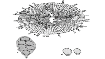
Figure 1: Vishnumyces malabarensis sp. nov. a. Appressoriate mycelium, b. Thyriothecium, c. Ascus, d. Ascospores
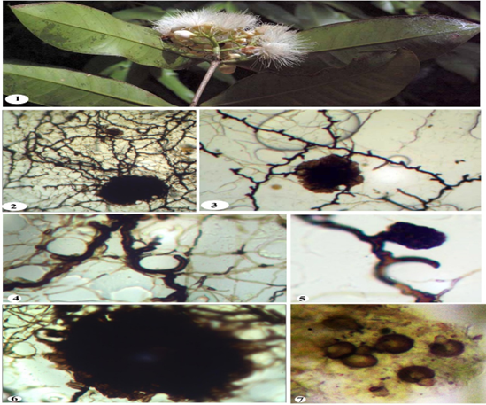
Plate-1: Vishnumyces malabarensis sp. nov.
1. Infected leaves of Syzygium mundagam (Bourd.) Chitra (Myrtaceae, 2. Colony with thyriothecia, 3. Branched appressoriate mycelium with thyriothecia, 4. Mycelium showing appressoria plugged around the stomata, 5. Developing thyriothecium, 6, Mature thyriothecium, 7. Ascospore
Colonies hypophyllous, subdense to dense, crustose, up to 2 mm in diameter, confluent. Hyphae substraight to crooked, branching opposite to irregular at acute to wide angles, loosely to rarely closely reticulate, cells 18-33 x 3-4 µm. Appressoria alternate to unilateral, antrorse to retrorse, ovate to clavate, curved to hook shaped, slightly truncate at the tip, 12-20 x 4-6 µm. Thyriothecia scattered, orbicular, stellately dehisced at the centre, up to 200 µm in diam., margin fimbriate, fringed hyphae straight to flexuous; asci oblong to cylindrical, slightly stipitate, bitunicate, octosporous, 40-60 x 30-40 µm; ascospores pale brown, uniseptate, septum at the distal end makes the spore proper “budding-off or pinching off” appearance, constricted at the septum to form one larger and one very smaller cell, 12-16 x 3-10 µm; larger cell ovate to oblong, 10-14 x 3-10 µm, smaller cell ovate to globose, often mammiform, 5-7 x 4-7 µm, wall smooth.
Material examined: On leaves of Syzygium mundagam (Bourd.) Chitra (Myrtaceae), Urakuzhy, Malabar Wildlife Sanctuary, Calicut, Kerala, India, Jan. 27, 2013, Lini K. Mathew, MTCHT 102 (Type), TBGT 6981 (isotype)
Etmology: The specific epithet is based Type locality.
Discussion
The genus Vishnumyces is reported here for the first time on the members of the family Myratceae (Hosagoudar and Harish, 2010). It is significant to that the ascospores germinate from the apical tip of the smaller cell by dissolving the cell wall and producing the mycelium from the inner content. Microscopic study revealed that the fungus possesses superficial mycelium and laterally located appressoria. The upper layer of the thyriothecia possessing radially arranged cells and dehiscing stellately at the centre. These are the characters of the family Asterinaceae and of the genus Asterina (Arx and Muller, 1975). Nonetheless, the septation at the terminal end of the ascospores results in variations in the size and shape of both cells. Hence, it has been accommodated in the genus Vishnumyces. The members of Asterinaceae showing host specificity and earlier, Vishnumyces was reported only on the host Otonephelium stipulaceum of Sapindaceae from Vazhachal forest, Trissur, Kerala (Hosagoudar et al, 2011; Hosagoudar, 2012). Hence based on the host specificity the present species may accommodated as a new species of the Genus Vishnumyces.
Based on the host specificity, colony morphology, measurement of mycelium and ascospores, shape, the present species can be included under the category of new species.
Declarations
Acknowledgement
I am grateful to Principal and HOD Botany, Mar Thoma College, Tiruvalla; KSCSTE, Govt. of Kerala for the facilities.
Funding Declaration
There was no funding for the above research work.
References
- Arx, J. A. V., & Müller, E. M. (1975). A re-evaluation of the bitunicate Ascomycetes with keys to families and genera. Studies in Mycology, 9:1–159.
Publisher | Google Scholor - Gamble, J. S. (1915–1936). The flora of the Presidency of Madras. Adlard & Son Ltd.
Publisher | Google Scholor - Henry, A. N., Kumari, G. R., & Chithra, V. (1987). Flora of Tamil Nadu, India (Ser. 1, Vol. 2). Botanical Survey of India.
Publisher | Google Scholor - Hosagoudar, V. B., Thomas, J., & Agarwal, D. K. (2011). Four new asterinaceous members from Kerala, India. Taprobanica, 3:15–17.
Publisher | Google Scholor - Hosagoudar, V. B. (2012). Asterinales of India. Mycosphere, 2:617–852.
Publisher | Google Scholor - Hosagoudar, V. B., Chandra Prabha, A., & Agarwal, D. K. (2011). Asterinales of Kerala. Associated Publishing Company, 270 + Pl. 23.
Publisher | Google Scholor - Hosagoudar, V. B., & Harish, M. (2010). Vishnumyces, a new genus of the family Asterinaceae from India. Indian Phytopathology, 63:85–86.
Publisher | Google Scholor - Sasidharan, N. (2004). Biodiversity documentation for Kerala – Flowering plants, Part 6:178.
Publisher | Google Scholor


