

Case Report
Lhermitte-Duclos Disease as An Incidental Finding Following an Isolated Syncopal Episode: A Case Report and Literature Review
- José David Sabbag
- Nicolas Lozano
- Andrés Aristizabal
- Valeria Gómez-Gomez *
- Daniel Suárez Sucre
Sabbag Radiologists, Universidad de La Sabana, Barranquilla, Colombia.
*Corresponding Author: Valeria Gómez-Gomez, Sabbag Radiologists, Universidad de La Sabana, Barranquilla, Colombia.
Citation: Sabbag JD, Lozano N, Aristizabal A, Gómez-Gómez V, Sucre DS. (2026). Lhermitte-Duclos Disease as An Incidental Finding Following an Isolated Syncopal Episode: A Case Report and Literature Review, International Journal of Biomedical and Clinical Research, BioRes Scientia Publishers. 7(5):1-7. DOI: 10.59657/2997-6103.brs.26.157
Copyright: © 2026 Valeria Gómez-Gomez, this is an open-access article distributed under the terms of the Creative Commons Attribution License, which permits unrestricted use, distribution, and reproduction in any medium, provided the original author and source are credited.
Received: August 20, 2026 | Accepted: September 21, 2026 | Published: October 06, 2026
Abstract
Lhermitte-Duclos disease, also known as dysplastic cerebellar gangliocytoma, is a rare hamartomatous lesion classified as a World Health Organization (WHO) grade 1 glioneuronal tumor. Its clinical presentation is variable and depends on the mass effect within the posterior fossa, with headache, ataxia, and signs of increased intracranial pressure being the most common manifestations. Some patients remain completely asymptomatic, and the diagnosis is established as an incidental imaging finding. We present the case of a female patient with a history of a single syncopal episode in whom a cerebellar lesion was incidentally detected on non-contrast cranial computed tomography (CT). Further characterization with contrast-enhanced brain magnetic resonance imaging (MRI) and proton magnetic resonance spectroscopy (MRS) demonstrated findings highly suggestive of Lhermitte-Duclos disease. Given the absence of hydrocephalus, fourth ventricular involvement, or significant mass effect, conservative management with imaging follow-up was undertaken.
Keywords: lhermitte-duclos disease; dysplastic cerebellar gangliocytoma; cerebellum; magnetic resonance imaging; magnetic resonance spectroscopy
Introduction
Lhermitte-Duclos disease (LDD) is an exceptionally rare entity characterized by dysplastic hypertrophy of cerebellar ganglion cells [1]. It is considered a low-grade glioneuronal tumor and is frequently associated with Cowden syndrome due to mutations in the PTEN gene [2]. Its true incidence remains unknown because of its rarity. At present, the diagnosis is primarily based on imaging findings, as the characteristic features observed on magnetic resonance imaging are sufficiently distinctive that histopathological confirmation by biopsy is generally unnecessary [3,14,15].
Case Presentation
A female patient with no significant neurological history presented after experiencing an isolated syncopal episode. As part of the initial evaluation, a non-contrast cranial computed tomography (CT) scan was performed, which incidentally revealed an expansile cerebellar lesion. Further characterization was subsequently performed with gadolinium-enhanced brain magnetic resonance imaging (MRI) and proton magnetic resonance spectroscopy (MRS), allowing for an appropriate assessment of the lesion.
Non-Contrast Cranial Computed Tomography (CT)
An intra-axial lesion was identified within the left cerebellar hemisphere. The lesion is round in morphology and predominantly hypodense, with heterogeneous attenuation due to the presence of internal hyperdense components. It measures 40 × 37 × 46 mm (anteroposterior × transverse × craniocaudal). A surrounding hypodense rim consistent with vasogenic edema is present, producing mass effect with displacement of the right cerebellar hemisphere and distortion of the fourth ventricle, without causing critical narrowing or obstruction. Alteration of the normal cerebellar folial architecture is also observed.
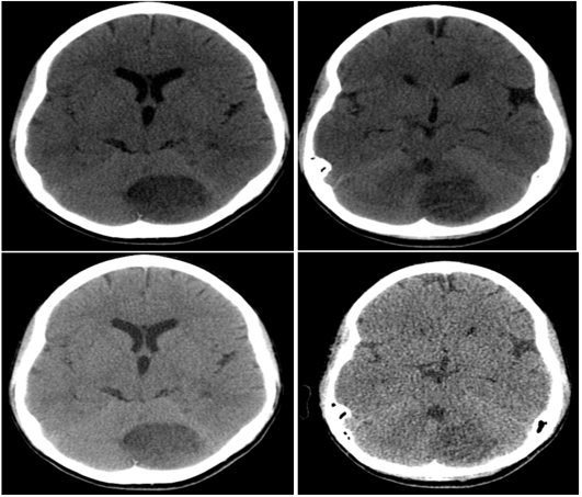
Figure 1: Computed tomography (CT): Cerebellar lesion. Low attenuation. Hemispheric thickening.
Key message: CT is usually nonspecific, and definitive diagnosis requires MRI.
Proton Magnetic Resonance Spectroscopy (MRS)
T2-weighted infratentorial signal abnormality involving the left cerebellar hemisphere, characterized by hyperintense signal within the cerebellar folial clefts, without displacement of the fourth ventricle, involving an area measuring approximately 45 mm in maximum diameter. An additional mixed hyperintense and hypointense subcortical supratentorial lesion was identified in the left cerebral hemisphere. The remaining cerebral white and gray matter demonstrated preserved differentiation. Quantitative and qualitative metabolic analysis showed normal levels of N-acetylaspartate (NAA), choline (Cho), creatine (Cr), and myo-inositol. A small lactate peak of no clinical significance was detected. No lipid peak was identified.
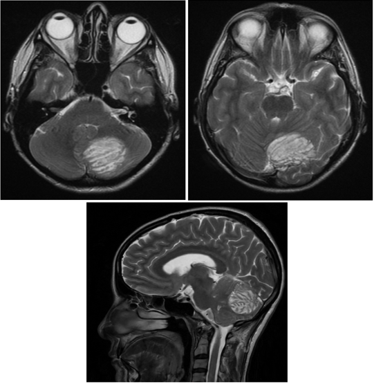
Figure 2: T2-weighted image: The striated pattern (“tiger stripe appearance”) observed on MRI is one of the most characteristic signs of this disease.
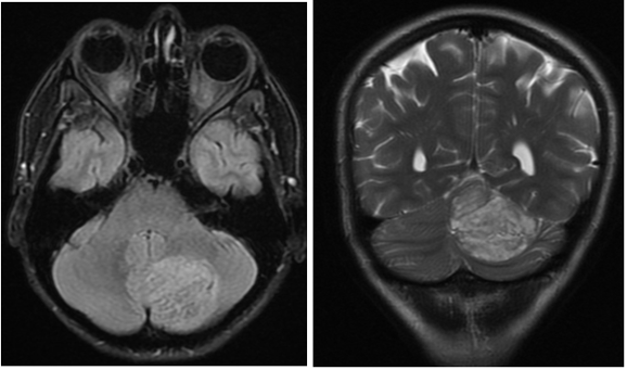
Figure 3: FLAIR- and T2 fat-saturated-weighted image: Striated pattern. Thickened folia.
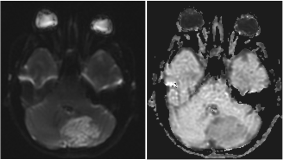
Figure 4: DWI-ADC: No marked diffusion restriction.
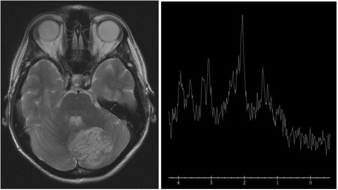
Figure 5: T2-weighted image: Laminar pattern. MOLECULAR: Compatible with a low-grade lesion, with no features suggestive of a high-grade tumor.
Contrast-Enhanced Magnetic Resonance Imaging (MRI)
A solid mass centered within the posteromedial aspect of the left cerebellar hemisphere was identified. The lesion is round in morphology, mildly hyperintense relative to the surrounding cerebellar parenchyma on T2-weighted images, and measures approximately 4.5 × 4.6 × 4.6 cm (craniocaudal × anteroposterior × transverse). The lesion demonstrates the characteristic "tiger-striped" appearance, with alternating bands corresponding to thickened cerebellar folia, and there is no associated vasogenic edema. Following intravenous gadolinium administration, several vascular structures are seen traversing the central portion of the lesion; however, the lesion itself demonstrates no significant contrast enhancement. Despite its relatively large size, there is no significant mass effect on the brainstem, nor is there obliteration of the fourth ventricle or the foramina of Luschka and Magendie.
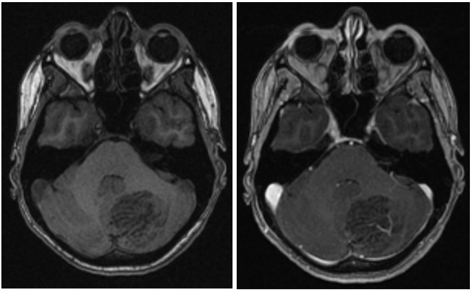
Figure 6: Non-contrast and contrast-enhanced: After intravenous contrast administration, some vascular structures are seen traversing the center of this lesion; however, the lesion itself does not show significant enhancement.
The imaging findings were considered highly suggestive of Lhermitte-Duclos disease. Given the absence of signs of increased intracranial pressure or cerebrospinal fluid (CSF) flow obstruction, the Neurosurgery Department elected to pursue conservative management with clinical and imaging follow-up. The patient was monitored for three consecutive years, during which no radiological or clinical changes were observed, further supporting the diagnosis.
Imaging Findings
Computed tomography (CT) is often the first imaging modality performed in patients with Lhermitte-Duclos disease; however, its findings are generally nonspecific. CT typically demonstrates a unilateral cerebellar lesion that is hypoattenuating or isoattenuating relative to the surrounding parenchyma, with little or no contrast enhancement and a variable degree of mass effect on the posterior fossa, depending on lesion size.
In contrast, magnetic resonance imaging (MRI) is the diagnostic modality of choice because of its high sensitivity for identifying the characteristic imaging features of the disease. The most distinctive finding is enlargement of the cerebellar folia, producing a laminated or striated appearance, commonly referred to as the "tiger-striped" or "corduroy" pattern. This appearance results from alternating hyperintense and iso- to hypointense bands on T2-weighted and FLAIR images, whereas the lesion is typically hypo- to isointense on T1-weighted sequences. Contrast enhancement following gadolinium administration is absent or minimal, diffusion restriction is generally absent or only subtle, and perfusion is usually normal or mildly increased, reflecting the low-grade nature of the lesion [2,7,11,19].
Magnetic resonance spectroscopy (MRS) provides complementary metabolic information and typically demonstrates a reduced N-acetylaspartate (NAA) peak, consistent with neuronal dysfunction, together with mild to moderate elevation of choline (Cho) related to cellular remodeling and a mildly increased choline-to-creatine ratio. A lactate peak may be present but is variable and is not considered a consistent finding. The combination of these imaging characteristics allows a highly confident diagnosis of Lhermitte-Duclos disease and facilitates differentiation from other cerebellar tumors and lesions, often eliminating the need for histopathological confirmation when the clinical and imaging findings are concordant [7,11].
Discussion
Lhermitte-Duclos disease is an exceedingly rare lesion whose pathophysiology is characterized by a developmental abnormality of the cerebellar cortex, resulting in hypertrophy of ganglion cells and disruption of the normal cerebellar architecture. Although some patients present with headache, ataxia, vertigo, seizures, or signs of increased intracranial pressure, others remain asymptomatic for many years [3,4,10].
Magnetic resonance imaging is the diagnostic modality of choice. The classic imaging feature is the characteristic "tiger-striped" appearance, which results from thickening of the cerebellar folia. Magnetic resonance spectroscopy provides useful metabolic information, generally demonstrating findings consistent with a low-grade lesion, although the metabolic profile may vary among patients.
The differential diagnosis includes cerebellar astrocytoma, medulloblastoma, hemangioblastoma, metastatic disease, subacute cerebellar infarction, and cerebellar cortical dysplasia. However, each of these entities has distinct imaging, pathological, and epidemiological characteristics that allow differentiation through clinicoradiological correlation, as summarized in Table 1 [2,7,11].
Table 1: Imaging features and differential diagnosis of Lhermitte-Duclos disease.
| Diagnosis | Epidemiology | CT Findings | MRI Findings | Distinguishing Features |
| Lhermitte-Duclos Disease | Rare. Typically affects young and middle-aged adults (30-50 years). May be associated with Cowden syndrome (PTEN mutation). | Hypoattenuating or isoattenuating cerebellar lesion with minimal mass effect. Calcifications are uncommon. | Characteristic laminated or striated ("tiger-striped" or "corduroy") appearance on T2-weighted and FLAIR images, with enlargement of the cerebellar folia; hypo- to isointense on T1-weighted images; absent or minimal diffusion restriction; absent or minimal contrast enhancement; low or mildly increased perfusion. MRS: decreased N-acetylaspartate (NAA), mild choline elevation, and variable lactate peak. | The cerebellar laminated ("tiger-striped") pattern is highly characteristic and considered virtually pathognomonic. |
| Pilocytic Cerebellar Astrocytoma | Most common in children and adolescents; uncommon in adults. | Predominantly cystic lesion with an isoattenuating or hyperattenuating mural nodule. | Cystic lesion with an intensely enhancing mural nodule; hyperintense on T2-weighted images; increased perfusion within the solid component. | The presence of a vividly enhancing mural nodule distinguishes it from Lhermitte-Duclos disease. |
| Medulloblastoma | Predominantly occurs in children; uncommon in adults. | Hyperattenuating midline or cerebellar hemispheric mass. | Hypointense on T1-weighted images, iso- to hypointense on T2-weighted images, marked diffusion restriction, variable contrast enhancement, and high cellularity. | Marked diffusion restriction and typical vermian location favor medulloblastoma. |
| Hemangioblastoma | Adults aged 30-60 years. May be associated with Von Hippel-Lindau syndrome. | Cystic lesion with a mural nodule and prominent vascular structures. | Intensely enhancing mural nodule with flow voids, markedly increased perfusion, and variable surrounding edema. | Marked hypervascularity and prominent feeding vessels are characteristic. |
| Cerebellar Metastasis | Older adults, usually with a history of primary malignancy (lung, breast, melanoma, or renal carcinoma). | Hypoattenuating lesion with vasogenic edema; hemorrhage or calcification may be present depending on the primary tumor. | Strong contrast enhancement (solid or ring-enhancing), prominent vasogenic edema, increased perfusion, and frequently multiple lesions. | Multiple lesions, disproportionate vasogenic edema, and a known primary malignancy strongly support the diagnosis. |
| Subacute Cerebellar Infarction | Patients with cardiovascular risk factors. | Territorial hypoattenuation following a vascular distribution. | Diffusion restriction during the acute phase; cortical enhancement and edema may develop during the subacute stage. Lesions conform to an arterial vascular territory. | Vascular distribution and temporal evolution distinguish infarction from Lhermitte-Duclos disease. |
| Cerebellar Cortical Dysplasia | Congenital disorder, usually diagnosed during childhood. | Nonspecific findings or subtle morphological abnormalities. | Disorganized cerebellar architecture with abnormal folial morphology and focal reduction or enlargement of cerebellar volume, without the characteristic laminated ("tiger-striped") appearance of Lhermitte-Duclos disease. | Developmental malformation without progressive mass effect or the characteristic laminated imaging pattern. |
Treatment
Management depends on the patient's clinical presentation. Surgical resection is indicated in patients with significant mass effect, hydrocephalus, or progressive neurological deterioration. In contrast, conservative management with periodic clinical and imaging follow-up is an accepted strategy for asymptomatic patients or those in whom the lesion is discovered incidentally [2,7].
Because of the well-established association between Lhermitte-Duclos disease and Cowden syndrome, clinical and genetic evaluation for PTEN mutations should be considered, particularly in patients with suggestive personal or family histories [5,6,10,12].
Conclusion
Lhermitte-Duclos disease is a rare entity that can be diagnosed with a high degree of confidence using magnetic resonance imaging, particularly when advanced techniques such as magnetic resonance spectroscopy are incorporated. Recognition of its characteristic imaging features may obviate the need for unnecessary invasive procedures. This case highlights the importance of careful evaluation of incidental findings on neuroimaging and demonstrates that even relatively large cerebellar lesions can be managed conservatively in the absence of fourth ventricular compromise, hydrocephalus, or significant mass effect [2,18,19].
Learning Points
- Lhermitte-Duclos disease is a rare cause of a cerebellar mass lesion.
- MRI demonstrates a characteristic laminated "tiger-striped" appearance that is highly suggestive of the diagnosis.
- Magnetic resonance spectroscopy supports the low-grade nature of the lesion by demonstrating a characteristic metabolic profile.
- Not all patients require surgical treatment; conservative management is appropriate in selected asymptomatic cases.
- Patients should be evaluated for the possible association with Cowden syndrome and PTEN gene mutations.
References
- Louis, D. N., Perry, A., Wesseling, P., Brat, D. J., Cree, I. A., et al. (2021). The 2021 WHO Classification of Tumors of The Central Nervous System: A summary. Neuro-Oncology, 23(8):1231-1251.
Publisher | Google Scholor - Alanazi, A. I., Alanezi, T., Aljofan, Z. F., Alarabi, A., Elwatidy, S. (2023). Lhermitte-Duclos Disease: A Systematic Review. Surgical Neurology International, 14:351.
Publisher | Google Scholor - Nowak, D. A., Trost, H. A. (2002). Lhermitte-Duclos Disease (Dysplastic Cerebellar Gangliocytoma): A Malformation, Hamartoma or Neoplasm? Acta Neurologica Scandinavica, 105(3):137-145.
Publisher | Google Scholor - Abel, T. W., Baker, S. J., Fraser, M. M., Tihan, T., Nelson, J. S., et al. (2005). Lhermitte-Duclos disease: A report of 31 cases with immunohistochemical analysis of the PTEN/AKT/mTOR pathway. Journal of Neuropathology & Experimental Neurology, 64(4):341-349.
Publisher | Google Scholor - Pérez-Núñez, A., Lagares, A., Benítez, J., Urioste, M., Lobato, R. D., et al. (2004). Lhermitte-Duclos disease and Cowden disease: Clinical and genetic study in five patients with Lhermitte-Duclos disease and literature review. Acta Neurochirurgica, 146(7):679-690.
Publisher | Google Scholor - Pilarski, R., Burt, R., Kohlman, W., Pho, L., Shannon, K. M., et al. (2013). Cowden Syndrome and The PTEN Hamartoma Tumor Syndrome: Systematic Review and Revised Diagnostic Criteria. Journal of the National Cancer Institute, 105(21):1607-1616.
Publisher | Google Scholor - Ma, J., Jia, G., Chen, S., Jia, W. (2019). Clinical Perspective on Dysplastic Gangliocytoma of The Cerebellum (Lhermitte-Duclos Disease). World Neurosurgery, 122:16-23.
Publisher | Google Scholor - Kumar, R., Vaid, V. K., Kalra, S. K. (2007). Lhermitte-Duclos Disease. Child's Nervous System, 23(7):729-732.
Publisher | Google Scholor - Ashraf, M., Kamboh, U. A., Raza, M. A., Choudhary, N., Mehboob, M., et al. (2022). Lhermitte-Duclos Disease: A Rare Cerebellar Hamartoma Presenting Following Traumatic Brain Injury and A Review of The Literature. Journal of Ayub Medical College Abbottabad, 34(3 Suppl 1):S733-S738.
Publisher | Google Scholor - Dragoo, D. D., Taher, A., Wong, V. K., Elsaiey, A., Consul, N., et al. (2021). PTEN Hamartoma Tumor Syndrome/Cowden Syndrome: Genomics, Oncogenesis, and Imaging Review for Associated Lesions and Malignancy. Cancers, 13(13):3120.
Publisher | Google Scholor - Joo, G., Doumanian, J. (2020). Radiographic Findings of Dysplastic Cerebellar Gangliocytoma (Lhermitte-Duclos Disease) in A Woman with Cowden Syndrome: A Case Study and Literature Review. Journal of Radiology Case Reports, 14(3):1-6.
Publisher | Google Scholor - Magaña, M., Landeta-Sa, A. P., López-Flores, Y. (2022). Cowden Disease: A Review. The American Journal of Dermatopathology, 44(10):705-717.
Publisher | Google Scholor - Zhou, X.P., Marsh, D. J., Morrison, C. D., Chaudhury, A. R., Maxwell, M., et al. (2003). Germline Inactivation of PTEN And Dysregulation of The Phosphoinositol-3-Kinase/Akt Pathway Cause Human Lhermitte-Duclos Disease in Adults. American Journal of Human Genetics, 73(5):1191-1198.
Publisher | Google Scholor - Padberg, G. W., Schot, J. D. L., Vielvoye, G. J., Bots, G. T. A. M., de Beer, F. C. (1991). Lhermitte-Duclos Disease and Cowden Disease: A Single Phakomatosis. Annals of Neurology, 29(5):517-523.
Publisher | Google Scholor - Robinson, S., Cohen, A. R. (2000). Cowden Disease and Lhermitte-Duclos Disease: Characterization of A New Phakomatosis. Neurosurgery, 46(2):371-383.
Publisher | Google Scholor - Borni, M., Kammoun, B., Kolsi, F., Abdelmouleh, S., Boudawara, M. Z. (2019). The Lhermitte-Duclos Disease: A Rare Bilateral Cerebellar Location of a Rare Pathology. Pan African Medical Journal, 33:118.
Publisher | Google Scholor - Bozbuga, M., Gulec, I., Suslu, H. T., Bayindir, C. (2010). Bilateral Lhermitte-Duclos Disease. Neurology India, 58(2):309-311.
Publisher | Google Scholor - García-Iturbide, R., Velázquez, J. A., Lozano Guzmán, I., Falcón-Molina, J. E., Rodríguez, M. A., et al. (2024). Treatment and Diagnostic Approach for Lhermitte-Duclos Disease and Suspected Cowden Syndrome. Cureus, 16(6):e62968.
Publisher | Google Scholor - Monjarás-Romo, G., Zavala-Romero, L., Tejada-Pineda, M. F., Meraz-Soto, J. M., Ballesteros-Herrera, D., et al. (2023). Lhermitte-Duclos Disease: A Case Series. Cureus, 15(8):e44326.
Publisher | Google Scholor - McMahon, M. E., Murray, D., MacNally, S., O’Brien, D. F. (2024). Lhermitte-Duclos Disease (Dysplastic Cerebellar Gangliocytoma) in The Setting of Cowden Syndrome: A Case Report and Literature Review on COLD Syndrome. British Journal of Neurosurgery, 38(5):1170-1173.
Publisher | Google Scholor