

Review Article
Features of the Application of Methods of Electroretinography and Tonometry of Intraocular Pressure to Assess the State of the Visual System of Children
1National Technical University of Ukraine Igor Sikorsky Kyiv Polytechnic Institute, Kyiv, Ukraine.
2Ternopil Ivan Puluj National Technical University, Ternopil, Ukraine.
*Corresponding Author: О.P. Yanenko, National Technical University of Ukraine Igor Sikorsky Kyiv Polytechnic Institute, Kyiv, Ukraine.
Citation: Yanenko O.P., Tkachuk R.M., Tkachuk R.A. (2026). Features of the Application of Methods of Electroretinography and Tonometry of Intraocular Pressure to Assess the State of the Visual System of Children, Clinical Case Reports and Studies, BioRes Scientia Publishers. 12(3):1-6. DOI: 10.59657/2837-2565.brs.26.311
Copyright: © 2026 O.P. Yanenko, this is an open-access article distributed under the terms of the Creative Commons Attribution License, which permits unrestricted use, distribution, and reproduction in any medium, provided the original author and source are credited.
Received: February 23, 2026 | Accepted: March 09, 2026 | Published: March 19, 2026
Abstract
The article discusses the features of creating a prototype system for parametric identification of electroretinal signal (ERS) and intraocular pressure (IOP) control for the purpose of reliable assessment and condition of the visual system in childhood, including the impact of neurotoxicity (identification of neurotoxicants, dose, characteristics and duration of exposure) range of daily deviation of changes in intraocular pressure.An analysis of the possibilities of using various optimization methods for parametric identification of the ERS model with low stimulation intensity was conducted.Special attention is paid to comparing the ERS processing time by known methods, analyzing their time complexity and studying the convergence of algorithms, reducing the duration of signal processing in the MATLAB software environment in order to obtain results in real time.Studies show that the use of effective optimization methods can significantly improve the accuracy (up to 5%) and speed of identification of ERS and IOP parameters(up to 12%-15%), which is critically important for reliable detection of the impact and prevention of negative health consequences already in the early stages of the disease.
Keywords: electroretinal signal; low intensity; neurotoxicity; intraocular pressure; processing optimization; parameter identification
Introduction
For modern studies of early stages of eye diseases and the state of the visual analyzer, it is important to have initial reliable information to identify the causes of visual impairment [1].The applied method of electroretinography (ERG) is of particular importance in studies of retinal disorders, since it is able to form an electro retinal signal (ERS), which indicates the high-quality functioning of rods and cones, and this ensures reliable diagnosis of the functions of the visual analyzer during monitoring [1,2].For example, in diseases such as retinitis pigmentosa, certain genetic disorders that lead to progressive degeneration of the retina, this method can detect a functional decrease in the response of photoreceptors even before the appearance of clinical symptoms. Similarly, with high intraocular pressure of the eye, which is subsequently characterized by blocking functions and damage to the optic nerve, it is necessary to monitor the rate of leakage of intraocular fluid. Also, recording ERG parameters can help assess the functional integrity of retinal ganglion cells, which are affected in the early stages of the disease, by measuring the electrical signals (responses) of different cell layers. Also, in the detection of diabetic retinopathy, a common complication affecting the blood vessels of the retina and is an important addition for the rapid and accurate assessment of the toxicity of certain drugs in the early stages of their use, as well as to detect the blocking of functional processes and the reduction of visual functions by some nanoparticles [3-5].
The development of networked computer technologies and digital analysis of response signals to various types of stimulation has significantly expanded the capabilities of low-intensity ERG. Modern electroretinographic systems already use advanced stimulation based on LED matrices to provide controlled light flashes of a given wavelength [5]. This approach ensures the creation of reproducible testing conditions, which is a necessary condition for increasing more accurate diagnostics in rapid studies.
The development of network computer technologies and digital analysis of response signals to various types of stimulation significantly expands the capabilities of low-intensity ERG. Modern electroretinographic systems already use advanced stimulation based on LED matrices to provide controlled light flashes of a given wavelength [5]. This approach provides the creation of reproducible testing conditions, which is a necessary condition for increasing more accurate diagnostics in rapid studies. Digital methods of processing the obtained results have made it possible to automate data recording, which accelerates obtaining accurate measurements of the retinal response, changes in morphological parameters (amplitudes and time components) and their specificity in changing the shape as a result of the effect of stimulation on photoreceptors (rods and cones), bipolar and ganglion cells. The signal selection technique involves the original placement of electrodes on the cornea of the eye to detect electrical activity generated by the retina in response to low light stimulation. This activity is recorded in a waveform that reflects the functional role of different layers of retinal cells. The importance of this method lies in the periodic testing of ERG, so ophthalmologists can monitor retinal function in real time, which allows timely prevention of deterioration of visual functions. Importantly, ERG and IOP changes also play a crucial role in the selection of new drugs with potential side effects of their effects on the retina and IOP, revealing neurotoxic effects already at the initial stage of action.
Research on optimization methods for determining the parameters of the test and real electro retinal signal in a model for detecting neurotoxicity.
It is known that the specificity of electro retinal signal processing, especially at ultra-low intensities under conditions of light stimulation, causes some complications in ensuring the accuracy and reliability of the results. Considering the signal-to-noise ratio in ERG, which can be insignificant at low levels of illumination. This means that the real signal from the retina can be noisy with background electrical activity under the influence of other sources, which makes it difficult to isolate important informative parameters of ERG and due to this influence their analysis and interpretation are complicated. The presence of artifacts caused by eye movement, blinking or minor shifts in electrode positioning can worsen the stability of the ERG signal and create prerequisites for making possible conclusions for their identification. The way out of this situation is the introduction of advanced methods of filtering and processing of signals and the obtained data before their analysis [6]. In turn, taking into account all components require advanced mathematical and computational procedures and tools to differentiate the signal into its constituent parts and more accurately assign them to specific categories in pathological conditions. The difference in ERG responses between individuals of different categories adds another layer of complexity. Factors such as age, individual retinal parameters and general health can cause deviations in ERG indicators, which requires the establishment of a range of reference values. This interindividual feature confirms the need for registration of large databases and reliable models and algorithms that can take these differences into account with the extraction of reliable diagnostic information [5-8]. Below (Figure. 1 a, b), a flowchart of the improved EMF processing and a fragment of the noisy electroretinogram before its optimal processing are presented.
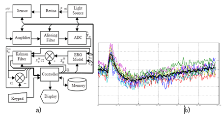
Figure 1a, b: Basic block diagram of EMF processing and Fragment of noisy electroretinogram before its processing: on the ordinate axis, the division value is 100 μV, on the abscissa axis – 50 ms.
Therefore, the creation of an improved electroretinographic system for new operating conditions requires refinement of the parameters of the EMF mathematical model and calculation algorithms. The test can include a larger number of data values, and their analysis requires powerful computing resources and improved calculation algorithms with real accuracy, which subsequently allow solving the tasks set.
In this work, an improved mathematical model of ERG using the Kalman filter is proposed and the principles of building a prototype of an information and measuring system for studying changes in the functional state of the retina at the early stages of detecting neurotoxicity are developed. These results are aimed at improving the diagnostic utility of the approach by increasing accuracy, reliability and reliable interpretation. Refinement of additional parameters of the mathematical model (namely, the coefficients of the difference equation) based on the use of the brute force method, which requires the necessary accuracy and convergence and significant calculation time. Reducing the duration of ERG data processing at low intensity of irritation allows you to reduce the time of monitoring the body in external cases. In the future, it is planned to use machine learning and artificial intelligence, offering potential solutions to speed up the process of data analysis and interpretation. Especially in the context of complex pathological conditions or minor functional changes, such as those occurring in the early stages of neurotoxicity, where a deep understanding of both the physiological processes in the retina and the mechanisms of their correlation with the results obtained is required.
In this case, studies have used advanced algorithms such as the Hooke-Jeeves method and the conjugate gradient method to accelerate the parametric identification of the electroretinographic signal [8]. The Hooke-Jeeves method is known for its simplicity and efficiency, especially in situations where the function does not have an analytic derivative or is non-differentiable. The method involves moving to find the minimum of the function by gradually refining the search direction. However, its efficiency and computational complexity depend significantly on the initial approximation conditions [8,9]. The conjugate gradient method is a more efficient algorithm for minimizing quadratic functions in nonlinear spaces. It iteratively updates the search direction, using gradient information to accelerate convergence. Despite its efficiency, it has certain limitations: the efficiency and time cost depend on the initial approximation conditions, and additional steps are required to effectively handle the initial constraints. Among the proposed methods are the Broyden-Fletcher-Goldfarb-Shannot (BFGS) method and the Nelder-Mead method.
The BFGS method is iterative, using an algorithm designed to minimize functions with nonlinear constraints. The main idea of the method is to approximate the Hessian matrix (which estimates the second derivatives of the functions) using a quasi-Newton approach. This approximation significantly reduces the computational burden compared to exact Hessian calculations. However, the BFGS method also has its own peculiarities. Storing the Hessian matrix approximation at each iteration can require a significant amount of memory, and the choice and definition of the initial approximation can significantly affect the speed and convergence of the method over time [9,10].
The Hessian matrix update rule Hk for the BFGS method at iteration k is given as:
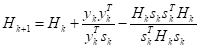 (1)
(1)
were
 (2)
(2)
and
 (3)
(3)
The Nelder-Mead perspective method is one of the most common for optimization. It works and is modified at each iteration to find the minimum or maximum of a function. The main idea of the Nelder-Mead method is to gradually expand or contract the simplex in the direction of optimization based on comparisons of functions at different points.
The Nelder-Mead algorithm involves several steps: mapping, expansion, compression, and reduction. These steps are used to transform a simplex in the function landscape in order to converge to an optimal solution. For example, the mapping step is mathematically represented as:
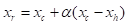 (4)
(4)
Where xt is the centroid of the simplex without the longest point xh, and α is the reflection coefficient. The convergence of optimization algorithms such as Nelder-Mead and BFGS can be determined by several criteria:
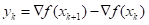
a) gradient norm – for methods that use derivatives, such as BFGS, convergence can be estimated by the gradient norm. If | |is below a set threshold, the algorithm is considered convergent.
b) Function value – convergence can also be assessed by observing changes in the function value. If the difference |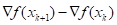 |is below a certain acceptable deviation, the algorithm is considered convergent.
c) Parameter change – In derivative-free methods like Nelder-Mead, convergence can be an indicator of parameter changes. If the simplex becomes small enough, such that || ||decreases below a certain threshold for all points in the simplex, convergence is said to be achieved.
The convergence of these algorithms depends on the nature of the objective function, initial conditions, and specific constraints of the problem. The correct choice of convergence criteria and consideration of the context of the problem allows maximizing the efficiency of the optimization process.
The BFGS method is used as an algorithm for minimizing functions subject to nonlinear constraints. It is based on the idea of approximating the quasi-Newton Hessian matrix, which evaluates the second derivatives of functions. However, the BFGS method also has its drawbacks: storing the Hessian matrix at each iteration can require significant memory, and the choice and definition of the initial approximation can significantly affect the speed and convergence of the applied method.
On the other hand, the Nelder-Mead method is one of the most widely used non-derivative optimization methods. It uses an iterative process where a set of points, called a simplex, is modified at each iteration to find the minimum or maximum of a function [11].The idea of the Nelder-Mead method is to gradually expand or contract the simplex towards the optimization direction based on comparisons of the function values at different points. Estimating the time and hardware complexity helps to determine the suitability of the BFGS method for a particular task and to identify limitations that may arise when using it for large-scale computing or real-time processing. Estimating the time and hardware complexity of optimization algorithms such as the Nelder-Mead method includes several aspects:
a) time complexity – the Nelder-Mead method has the features of being adaptive and having no explicit gradients. However, it is often used as a method with a worse time complexity of O(n2), where n is the number of parameters or the dimension of the parameter space. This method iteratively adapts a simplex (a geometric figure in the parameter space) to minimize or maximize the objective function. Each iteration involves evaluating the function at several points on the simplex, mapping, expanding, or contracting it depending on the function estimates, which contributes to the overall time complexity.
b) hardware complexity – similar to the BFGS method, the Nelder-Mead method does not require specialized hardware and can be run on conventional computing devices. However, the memory requirements may vary depending on the size of the parameter space and the number of iterations required for convergence. The memory usage of the method involves mainly storing the vertices of the simplex and function estimates, which can increase with increasing size of the parameter space.
c) Memory – The memory requirements for the Nelder-Mead method depend mainly on the dimensionality of the parameter space and the number of iterations. Although the method does not explicitly compute gradients or Hessian matrices, it still needs to store function estimates and simplex vertices. As the number of parameters increases, memory usage can increase, which can be a limiting factor for large-scale optimization problems.
Estimating the time and hardware complexity of the Nelder-Mead method helps to assess its suitability for specific optimization problems and provides an idea of the resource requirements, which allows making informed decisions about its application for various computational tasks [8-10].
Determining the convergence of optimization algorithms for the Nelder-Mead and BFGS methods usually involves monitoring certain criteria during algorithm iterations. Therefore, it is possible to estimate the convergence for each of the methods:
a). objective function value – tracking the value of the function at each iteration. If the value of the function gradually decreases or stabilizes within a given accuracy, this indicates its convergence.
b). simplex compression – checking whether the simplex (the geometric shape that encompasses the current solution) is compressed to a minimum or maximum point. Convergence occurs when the simplex is compressed to a small size, indicating that it is approaching the optimum.
c). parameter variation – examining the change in parameter values from one iteration to the next. Convergence is achieved when the parameter values stabilize into a stable solution within a specified accuracy and convergence efficiency of the optimization process.
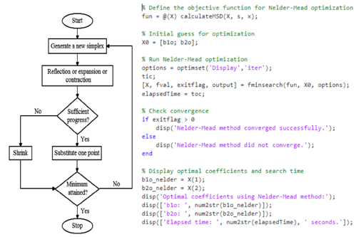
Figure 2: Flowchart of Nelder-Mead simplex algorithm and convergence determination in MATLAB environment
The most widely used of the derivative-free optimization methods, which is widely known in engineering, is the adapted Nelder-Mead algorithm shown above in the block diagram in Fig. 2. Its main advantage is its ability to handle functions that may obviously have noise present. This is the main reason for justifying the advantages of this algorithm and analyzing its convergence rate. The Nelder-Mead algorithm does not require the calculation of the derivatives of the function, which makes it suitable for optimizing functions where the calculation of derivatives is difficult or impossible. This is a significant advantage in problems where the function has a complex analytical form. The algorithm uses only the values of the functions to modify the simplex (in n-dimensional space), which makes it attractive for practical applications. The method is also applicable to a wide range of optimization problems, including those with multiple local minima (real ERG).Its flexibility allows it to adapt to different types of functions in multidimensional spaces, as it does not require the construction of global models. Since the method does not use derivatives, it requires less RAM and is less sensitive to noise in function values.
The dependence of the convergence rate of the Nelder-Mead algorithm on factors including the initial configuration of the simplex, the nature of the optimized function and the stopping criteria. The rate of approximation to the optimal solution increases linearly with the number of iterations. The time complexity of this algorithm depends on the dimension of the parameter space n. At each iteration, the method requires the evaluation of the function at n+1 points, which leads to the need to estimate the complexity O(n) per iteration. However, with the availability of high-performance computers and improved algorithms for processing the results of the study, the possibility of working in real time is achieved. Also, the use of an improved model for assessing the leakage of intraocular fluid in glaucoma significantly reduces the process of obtaining results about the state of the visual system in children at the initial stage of the disease. Therefore, the creation of a prototype system for parametric identification of the electroretinal signal (ERS) and control of intraocular pressure (IOP) in order to reliably assess the state of the visual system, including the impact of neurotoxicity in childhood, is a prospect of modern research.
Conclusion
1. The Nelder-Mead method is known for its versatility and reliability, ensuring the efficiency of optimizing complex, uneven or noisy objective functions. Its adaptability in various optimization conditions allowed creating an expert electroretinographic system for the tasks of identifying human neurotoxicity in real time.
2. The time complexity of the applied Nelder-Mead algorithm was evaluated and a 13-15% reduction in the time of determining the necessary parameters during the system operation in the test mode of electroretinal signal processing was established.
3. The use of the proposed Nelder-Mead algorithm provided 10-30% less computing power in the core, which in real time makes it possible to use it in prototypes of expert information and measurement systems for analyzing the parameters of the visual analyzer state.
4. An improved model for calculating the rate of intraocular fluid leakage for the initial stage of glaucoma development in children was also developed.
5. The main approaches to creating a prototype of a system for parametric identification of electroretinal signal (ERS) and monitoring of intraocular pressure (IOP) have been established for the purpose of reliable assessment of the state of the visual system in childhood.
References
- American Academy of Ophthalmology. (2024). Primary congenital glaucoma. EyeWiki.
Publisher | Google Scholor - Tkachuk, R. A. (2010). Research on human neurotoxicity by nanomaterials. In Proceedings of the International Scientific and Technical Conference Current Problems of Biomedical Engineering, Informatics, Cybernetics and Telemedicine (7). Kyiv, Ukraine: National Technical University of Ukraine Igor Sikorsky Kyiv Polytechnic Institute.
Publisher | Google Scholor - Cornish, E. E., Vaze, A., Jamieson, R. V., & Grigg, J. R. (2021). The electroretinogram in the genomic era: Outer retinal disorders. Eye, 35(12):2406–2418.
Publisher | Google Scholor - McCulloch, D. L., Marmor, M. F., Brigell, M. G., Hamilton, R., Holder, G. E., Tzekov, R., & International Society for Clinical Electrophysiology of Vision. (2015). ISCEV standard for full-field clinical electroretinography (2015 update). Documenta Ophthalmologica, 130(1):1–12.
Publisher | Google Scholor - Tatham, A. J., & Medeiros, F. A. (2017). Detecting structural progression in glaucoma with optical coherence tomography. Ophthalmology, 124(4 Suppl.):S57–S65.
Publisher | Google Scholor - Tkachuk, R. A., Yavorsky, B. I., & Yanenko, O. P. (2015). Evaluation of the risk of neurotoxicity with the help of electroretinography. Bulletin of the National Technical University of Ukraine Kyiv Polytechnic Institute. Series: Radio Engineering, Radio Equipment Construction, 61:108–115.
Publisher | Google Scholor - Robson, A. G., Nilsson, J., Li, S., Jalali, S., Fulton, A. B., Tormene, A. P., Holder, G. E., & Brodie, S. E. (2018). ISCEV guide to visual electrodiagnostic procedures. Documenta Ophthalmologica, 136(1):1–26.
Publisher | Google Scholor - Tymkiv, P. (2021). Analysis of the complexity of algorithms for finding the coefficients of the mathematical model of low-intensity electroretino signal. In Advanced Applied Energy and Information Technologies 2021: Proceedings of the International Conference (145–150). Ternopil, Ukraine.
Publisher | Google Scholor - Gill, M. A., & Miller, J. D. (2011). Muskingum models using Nelder–Mead simplex algorithm. Journal of Hydrologic Engineering, 16(11):946–954.
Publisher | Google Scholor - Nocedal, J., & Wright, S. J. (2016). Optimization methods: A review. SIAM Review, 58(3):492–525.
Publisher | Google Scholor - Tkachuk, R. M., et al. (2024). Modeling the process of stress formation in the eyeball in childhood glaucoma. In Proceedings of the 23rd International Scientific and Technical Conference Instrumentation: State and Prospects Kyiv, Ukraine: National Technical University of Ukraine Igor Sikorsky Kyiv Polytechnic Institute, (165–168).
Publisher | Google Scholor


