

Case Report
Breast Cancer: Performance of [68Ga] Ga-FAPI PET following Baseline [18F] F-FDG PET
1Ocean Road Cancer Institute, Dar es salaam, Tanzania.
2Department of Clinical Oncology, Muhimbili University of Health and Allied Sciences (MUHAS), Dar es Salaam, Tanzania.
*Corresponding Author: Bright Awadh Sangiwa, Ocean Road Cancer Institute, Dar es salaam, Tanzania.
Citation: Sangiwa B A, Sabaya F, Fundo B, Ramadhani S. (2025). Breast Cancer: Performance of [68Ga] Ga-FAPI PET following Baseline [18F] F-FDG PET. International Clinical Case Reports and Reviews, BioRes Scientia Publishers. 3(2):1-3. DOI: 10.59657/2993-0855.brs.25.034
Copyright: © 2025 Bright A. Sangiwa, this is an open-access article distributed under the terms of the Creative Commons Attribution License, which permits unrestricted use, distribution, and reproduction in any medium, provided the original author and source are credited.
Received: February 18, 2025 | Accepted: March 10, 2025 | Published: March 14, 2025
Abstract
A 59-Year-old woman, post menopause, presented with one year history of left breast mass of about 5cm associated with skin ulceration. The core biopsy was performed and revealed left breast invasive ductal carcinoma (IDC) grade II, ER +++, PR ++, Her ++, FISH neg, Ki-67: 16%.
Keywords: carcinoma; skin ulceration; metastasis; breast tumor
Introduction
Baseline [18F] F-FDG PET -CT scan was done in the light of confirmed biopsy to assess locoregional nodal and distant metastasis. The study demonstrated increased FDG uptake in the left breast mass with skin, posterior chest wall involvement, multiple small satellite lesions, bilateral axillary and retropectoral lymph nodes (Figure 1 and 2).
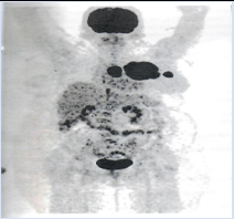
Figure 1: FDG PET MIP projection, three lesions were seen demonstrating intense FDG uptake in the chest.
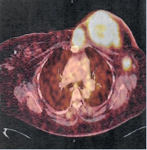
Figure 2: FDG PET-CT fusion axial projection showed intense FDG uptake in the left breast mass, a satellite mass abutting the sternum and retropectoral lymph node.
The option of treatment was disposed to the patient about chemotherapy versus hormonal therapy in neoadjuvant setting. The patient chose the latter and was started on Tabs Letrozole 2.5 mg once a day for 6 months and Tabs Ribociclib 600 mg for 21 days then 7 days off (every 28 days cycle for 4 cycles). Blood tests were done every after 14 days and were essentially normal throughout the treatment.
After the completion of neoadjuvant chemotherapy, the patient visited our center for follow up [18F] F-FDG PET -CT scan to assess treatment response, however at the time of her visit FDG PET was unavailable. Alternative imaging PET modality with [68Ga] Ga-FAPI PET-CT scan was explained to patient and his family.
[68Ga] Ga-FAPI PET-CT scan was then performed and the interpretation with comparison to baseline [18F] F-FDG PET -CT scan was done by the same user. The study demonstrated abnormal FAPI uptake in the known left breast tumor and one ipsilateral satellite small chest lesion, both with central areas of no uptake in keeping with necrotic tissue. On low dose CT, the breast tumour measured 5.1 x 5.8 cm (AP X TV) and a satellite mass abutting the sternum measured 2.1 x3 cm (AP x TV) with no underlying bone changes (Figure 3 & 4).
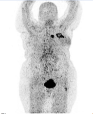
Figure 3: FAPI PET MIP projection showed two lesions in the chest with intense FAPI uptake, one with central area of no FAPI uptake.
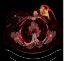
Figure 4: FAPI PET-CT fusion projection showed intense FAPI uptake in the left breast and a satellite mass, both with central necrosis. Intense FAPI uptake was also seen in the retropectoral lymph node located just below the left breast mass.
There was also a low-grade uptake involving retropectoral node. The left axillary lymph nodes also demonstrated low grade uptake with fat hila on CT, these were considered benign. On dedicated lung window CT, lung fields were clear (Figure 5).
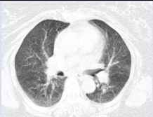
Figure 5: Dedicated chest CT showed lung fields were clear.
The patient was scheduled for surgery, left mastectomy with left axillary lymph nodes clearance was performed. The histopathology showed malignant breast tissue (IDC) with central necrosis and one metastatic lymph node (1/8), these findings concurred with what we reported on [68Ga] Ga-FAPI PET-CT. The patient was referred to an oncologist and currently undergoing chemo-radiation treatment.
Discussion
[18F] F-FDG PET-CT is still considered as a gold standard nuclear medicine PET imaging modality in breast cancer patient to assess response to treatment [1]. However, in recent years, there has been introduction of an alternative to [18F] F-FDG PET -CT with [68Ga] Ga-FAPI PET-CT scan in different cancers [2]. Literatures have also reported [68Ga] Ga-FAPI PET -CT performs better in the detection of primary breast tumour and distant metastasis (hepatic and brain) due to its low background activity [2,3]. The new practical guideline for FAPI PET has further demonstrated the useful of this modality in different cancers [4].
Our study has demonstrated the useful of [68Ga] Ga-FAPI PET-CT as an alternative PET modality for patient with baseline [18F] F-FDG PET-CT, when FDG PET is unavailable.
Learning point
[68Ga] Ga-FAPI PET-CT is an alternative PET modality in breast cancer patients when or where [18F] F-FDG PET-CT is not accessible.
[68Ga] Ga-FAPI PET-CT requires no patient preparation as compared to [18F] F-FDG PET-CT preparation of radiopharmaceutical does not require cyclotron.
Declarations
Funding: No funding
Competing interests: None declared.
Declaration of patient consent: Written consent for all images and clinical information was obtained.
References
- Sofia C. Vaz, John Patrick, Pilkington Woll, Fatima Cardoso, et al. (2024). Joint EANM‑SNMMI guideline on the role of 2‑[18F] FDG PET/CT in no special type breast cancer (endorsed by the ACR, ESSO, ESTRO, EUSOBI/ESR, and EUSOMA). Eur J Nucl Med Mol Imaging, 2706-2732.
Publisher | Google Scholor - Kratochwil C, Flechsig P, Lindner T, et al. (2019). 68Ga-FAPI PET/CT: Tracer uptake in 28 different kinds of cancer. J Nucl Med, 60:801-805.
Publisher | Google Scholor - Silvia Taralli, Margherita Lorusso, Elisabetta Perrone, et al. (2023). PET/CT with Fibroblast Activation Protein Inhibitors in Breast Cancer: Diagnostic and Theranostic Application: A Literature Review, Cancers, 15:908.
Publisher | Google Scholor - Hope A, Jeremie Calais, Ajit H. Goenka et al. (2024). SNMMI Procedure Standard/EANM Practice Guideline for Fibroblast Activation Protein (FAP) PET. Journal of Nuclear Medicine, 124.269002.
Publisher | Google Scholor


